10
A routine physical exam, a scan of your carotid arteries, or an imaging workup after a minor neck strain often brings an unexpected discovery: a thyroid nodule. Within hours, a notification pops up on your patient portal, presenting a dense radiology report filled with intimidating phrases like “hypoechoic,” “punctate calcifications,” or “TI-RADS 4.” For most people, reading those clinical descriptions triggers an immediate wave of anxiety, followed by an urgent impulse to search online forums to determine whether they might have thyroid cancer.
The reality is far more reassuring than a raw imaging report might make it seem. Thyroid nodules are extraordinarily common in the adult population. High-resolution ultrasound studies consistently show that well over half of all adults over the age of fifty have at least one thyroid nodule. More importantly, the vast majority of these nodules—over ninety to ninety-five percent—are completely benign.
An ultrasound exam does not deliver a final diagnosis of cancer. Instead, it functions as an acoustic map that helps physicians evaluate risk, decide whether a tissue sample is necessary, or confirm that simple monitoring is the safest course of action. Understanding how radiologists interpret these images can transform an overwhelming medical document into clear, actionable knowledge.
Why Ultrasound Is the Primary Tool for Thyroid Evaluation
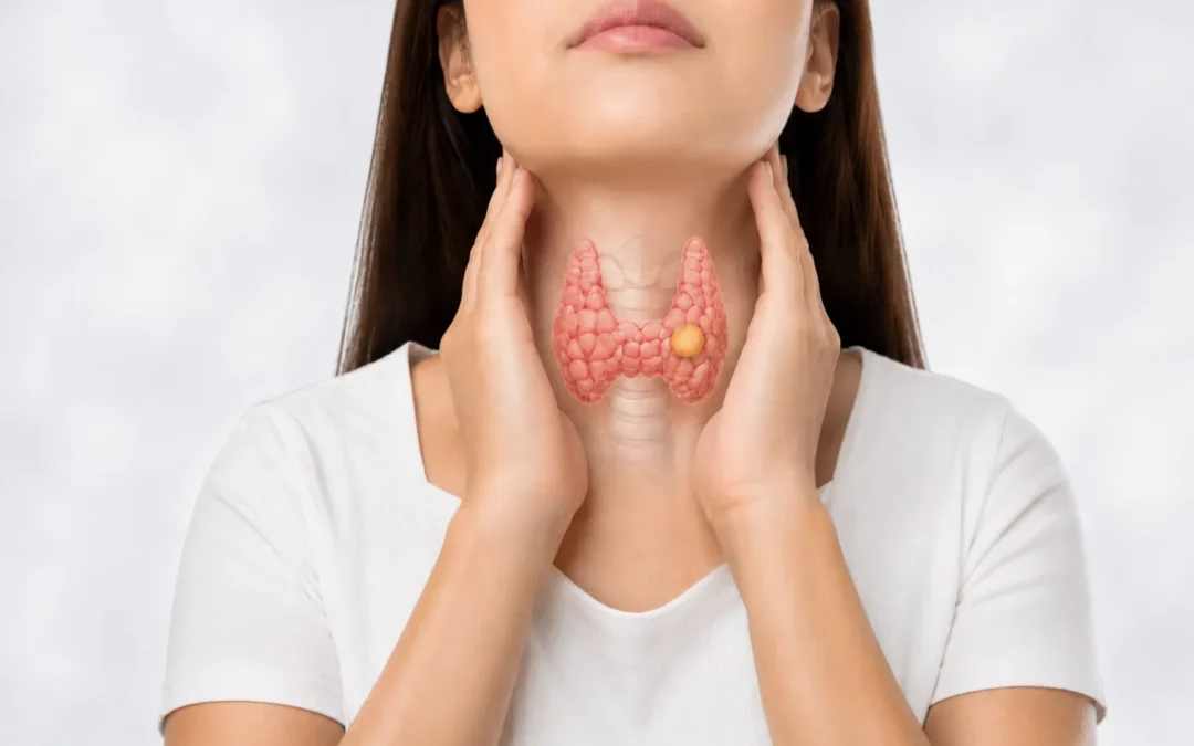
The thyroid is a small, butterfly-shaped endocrine gland situated at the base of your neck, resting just in front of the windpipe. Because of its superficial location right beneath the skin and a thin layer of muscle, it is uniquely suited for acoustic imaging. High-frequency sound waves can pass through the overlying tissues to generate detailed, high-resolution pictures of the gland in real time, all without exposing you to ionizing radiation.
While blood tests like thyroid-stimulating hormone (TSH) evaluate how well the gland is functioning biochemically, an ultrasound examines its physical architecture. It can distinguish between fluid and solid cellular material, measure dimensions down to the millimeter, and evaluate internal structures that cannot be felt during a physical examination.
Furthermore, sonographers can switch on Doppler ultrasound to observe blood flow patterns within and around a nodule, while simultaneously checking adjacent lymph nodes in the neck. This provides a comprehensive overview that helps doctors separate routine, harmless findings from those that warrant closer investigation.
Decoding the Vocabulary on Your Ultrasound Report
Radiologists use a standardized lexicon to describe the physical characteristics of thyroid nodules. When you review your report, you will see observations broken down into several structural categories: composition, echogenicity, borders, shape, and internal reflective patterns. Each attribute contributes a distinct piece of the diagnostic puzzle.
Composition: Fluid, Solid, or Spongiform
Composition describes the physical makeup of the nodule:
A purely cystic nodule is filled entirely with fluid. Because fluid allows sound waves to pass straight through without bouncing back, cysts appear pitch black on the screen. Pure cysts are virtually always benign and rarely require intervention unless they enlarge enough to cause local pressure or cosmetic concerns.
A spongiform nodule features a microcystic, honeycomb-like pattern where tiny fluid pockets are scattered throughout healthy tissue. This classic acoustic pattern is overwhelmingly benign, typically reflecting normal fluctuations in colloid tissue or long-standing hyperplastic growth.
A solid nodule consists entirely of cellular tissue. While the overwhelming majority of solid nodules are still benign, solid tissue is where abnormal cell growth occurs. As a result, solid nodules receive the closest scrutiny.
A mixed cystic and solid nodule contains both liquid and cellular components. When interpreting these, the radiologist focuses almost entirely on the appearance of the solid portion to estimate risk.
Echogenicity: How the Nodule Reflects Sound
Echogenicity describes the brightness of the nodule relative to the surrounding healthy thyroid tissue:
An anechoic structure reflects no sound waves, appearing dark black, which is characteristic of fluid.
An isoechoic nodule reflects sound at the same intensity as normal thyroid tissue, while a hyperechoic nodule appears brighter. Both findings are broadly reassuring and strongly associated with benign adenomas.
A hypoechoic nodule appears distinctly darker than the surrounding gland. Denser cellular clusters absorb and scatter sound waves differently, creating this darker shade. While many benign nodules are hypoechoic, the majority of thyroid malignancies also present this way, making it a feature that prompts careful documentation.
A markedly hypoechoic nodule appears darker than the strap muscles located just in front of the thyroid gland. This is a more notable finding that carries a higher statistical association with suspicious cellular changes.
Margins and Shape: Growth Patterns and Borders
The boundary where a nodule meets normal tissue reveals how it interacts with its surroundings:
A smooth, well-circumscribed margin shows that the nodule is expanding within its own capsule, respecting surrounding tissue planes. This is a hallmark of non-cancerous growth.
An irregular, lobulated, or ill-defined margin means the borders appear jagged or blurred, suggesting that the nodule may be pushing unevenly into adjacent thyroid tissue.
The term taller-than-wide describes a nodule whose front-to-back measurement exceeds its side-to-side measurement. Because benign nodules tend to grow horizontally along natural tissue planes, a nodule that grows vertically across tissue layers warrants greater caution.
Calcifications: Types of Calcium Deposits
Ultrasound waves reflect intensely off mineral deposits, showing up as bright white spots that sometimes cast dark acoustic shadows behind them:
Macrocalcifications are large, coarse mineral deposits often caused by past inflammation, minor tissue bleeding, or the natural aging of a long-standing nodule. They are generally not an indicator of aggressive disease.
Rim or eggshell calcifications form a thin, curved perimeter around the edge of a nodule. These are commonly benign, though radiologists will verify that the shell remains continuous and does not have soft tissue protruding through an opening.
Punctate echogenic foci, or microcalcifications, appear as tiny, pinprick-bright specks without significant acoustic shadowing. Within solid nodules, these can represent microscopic calcium bodies known as psammoma bodies, which are frequently linked with papillary thyroid carcinoma.
Understanding the TI-RADS Scoring System
To eliminate subjectivity and prevent conflicting interpretations between different clinics, professional medical associations established the Thyroid Imaging Reporting and Data System, commonly known as TI-RADS. In the United States, the American College of Radiology (ACR) TI-RADS model is the recognized standard.
Rather than relying on intuitive impressions, ACR TI-RADS assigns numerical points across five visual categories: composition, echogenicity, shape, margin, and echogenic foci. The points are added together to place the nodule into a risk tier:
TR1 (Benign, 0 points): Demonstrates essentially zero risk of malignancy.
TR2 (Not Suspicious, 2 points): Includes findings like spongiform nodules, carrying a cancer risk below two percent.
TR3 (Mildly Suspicious, 3 points): Represents a low-risk profile with a malignant probability around five percent.
TR4 (Moderately Suspicious, 4 to 6 points): Carries an intermediate risk, with malignant probabilities typically ranging between five and twenty percent.
TR5 (Highly Suspicious, 7 or more points): Reflects multiple atypical features, indicating a risk of malignancy greater than twenty percent.
The most critical takeaway is that a higher TI-RADS score does not automatically mean you need a biopsy. The system pairs the risk tier directly with the nodule’s physical size before recommending any invasive steps.
For instance, a TR3 nodule generally does not warrant a biopsy unless it reaches at least 2.5 centimeters. A TR4 nodule usually requires sampling only if it measures 1.5 centimeters or more. Even a TR5 nodule rarely undergoes biopsy if it is smaller than 1.0 centimeter.
These size thresholds exist to protect patients from overdiagnosis and overtreatment. Decades of clinical tracking have demonstrated that tiny, low-risk thyroid lesions—even those that might technically harbor microscopic papillary changes—rarely grow, spread, or impact health over an individual’s lifetime. By factoring size into every recommendation, TI-RADS helps physicians avoid subjecting patients to unnecessary procedures.
Why Size Does Not Equal Danger
A common source of confusion for patients is equating size with severity. It is intuitive to assume that a large growth must be dangerous, while a tiny one can be safely ignored. With the thyroid, that logic rarely holds true.
A benign colloid nodule or follicular adenoma can easily expand to three, four, or five centimeters across, creating a visible lump or a sensation of fullness in the throat, while remaining entirely harmless. Conversely, a malignant nodule might remain a dormant, four-millimeter speck that never causes a problem.
When an ultrasound report notes that a nodule measures two centimeters, that measurement reflects spatial volume, not cancer risk. Size becomes clinically meaningful in two scenarios: when evaluating whether the nodule crosses the biopsy threshold for its specific TI-RADS score, and when determining whether it is large enough to cause physical symptoms like swallowing difficulties, local pressure, or chronic throat clearing.
When Is a Biopsy Actually Recommended?
When a nodule displays suspicious characteristics and meets the corresponding size criteria, your physician will likely suggest a Fine-Needle Aspiration (FNA).
An FNA is a quick, straightforward outpatient procedure. After numbing the skin with a local anesthetic, the physician uses continuous ultrasound guidance to track the needle tip in real time, advancing an ultra-thin needle—often smaller than the needles used for standard blood draws—into the nodule to collect cellular samples. The entire process takes only a few minutes, involves minimal discomfort, and allows you to return to normal activities right away.
The harvested cells are sent to a cytopathology lab and evaluated using the Bethesda System for Reporting Thyroid Cytopathology. This framework sorts results into six categories:
Bethesda I (Nondiagnostic): The sample did not yield enough intact cells for a definitive read, meaning the procedure may need to be repeated.
Bethesda II (Benign): This represents approximately seventy percent of all thyroid biopsies, confirming a risk of malignancy lower than three percent.
Bethesda III and IV (Indeterminate): The cells display subtle irregularities that make it difficult to categorize them cleanly as benign or malignant. In modern clinical practice, these samples often undergo advanced molecular genetic testing, which checks for specific gene mutations and spares the vast majority of patients from exploratory surgery.
Bethesda V (Suspicious for Malignancy) and VI (Malignant): The sample contains distinctive markers of thyroid cancer, prompting a surgical consultation to discuss removing part or all of the thyroid gland.
Active Surveillance and Long-Term Monitoring
If your ultrasound indicates that a biopsy is unnecessary, the standard recommendation is periodic observation, often called active surveillance. For low-risk or smaller nodules, your doctor will usually schedule a follow-up ultrasound in twelve, eighteen, or twenty-four months.
The goal of monitoring is to verify stability over time. Benign nodules frequently fluctuate in size, expanding slowly or even shrinking as internal fluids absorb and resolve. Stability over consecutive imaging studies provides strong confirmation of benign behavior.
Radiologists compare previous scans against the latest images to assess interval growth, typically defined as an increase of at least twenty percent in two dimensions or a fifty percent increase in total volume. If a nodule shows no meaningful change across several follow-ups, the spacing between future ultrasounds can often be widened significantly.
Navigating Your Next Steps with Confidence
Reviewing an imaging report dense with technical terminology can feel alarming, but context is essential. Thyroid nodules are an expected finding on head and neck imaging, not a rare emergency. A radiologist’s role is to catalog every acoustic detail with meticulous precision, not to deliver a distressing diagnosis.
If your report suggests follow-up, arrange a visit with your primary care provider, an endocrinologist, or an ear, nose, and throat specialist. Bring your full written report and, if possible, the digital imaging disc so your physician can evaluate the images firsthand. In most instances, your appointment will conclude with a clear plan for routine monitoring rather than immediate intervention. Understanding the language of your ultrasound allows you to approach your results with confidence, clarity, and peace of mind.